Subtotal $0.00
Pseudocyst
Follows an attack of pancreatitis, and holds pancreatic juice rather than tumour tissue. Usually the collection settles without any procedure, though large or infected pseudocysts need drainage.

Specialist gastroenterology, liver and pancreas care in KPHB Colony, Kukatpally. Sunday OPD, evening hours and on-site FibroScan.
Pancreatic cystic lesions in KPHB need careful assessment rather than instant surgery. Generally, these cysts stay harmless for many years, but a small group carries a real cancer risk — so the correct test at the correct time matters far more than speed. At Gokul Gastro & Liver Care, Dr. Viswanath Kamisetty reviews every scan personally.
Sunday OPD, evening hours, on-site FibroScan, and a written surveillance plan with actual dates handed to you at every visit — under one DM-qualified specialist, start to finish.
Monday – Saturday
Sunday
A pancreatic cystic lesion is a fluid-filled pocket inside or beside the pancreas. Scans usually pick these up by accident — many patients reach us after an ultrasound ordered for something unrelated. The first question is never "operate or not"; instead, we ask what kind of cyst this actually is.
These lesions fall into two broad families. Some are collections left behind by inflammation; others are true tumours that grow very slowly. Because both look alike on a basic scan, detailed imaging becomes essential, and behaviour shifts with size, location and duct connection. Detection rises sharply after fifty, though younger adults do develop cysts too, particularly after pancreatitis.

Type decides everything that follows. In practice, six patterns appear regularly.
Follows an attack of pancreatitis, and holds pancreatic juice rather than tumour tissue. Usually the collection settles without any procedure, though large or infected pseudocysts need drainage.
Almost always benign, with a honeycomb pattern on scans, and more common in older women. Surveillance alone is usually enough — we act only when the cyst grows large and presses on nearby organs.
The lining produces mucin, so malignant change remains possible. Typically sits in the body or tail of the pancreas, usually in women in their forties and fifties, and surgeons often remove it because of that cancer potential.
Grows from the pancreatic duct lining itself, and risk depends heavily on which duct is involved. Main-duct IPMN carries high cancer risk, so specialists usually advise resection; branch-duct IPMN carries much lower risk, so many simply enter long-term surveillance.
A rare tumour, with young women forming the classic group. It looks alarming on first mention, but outcomes after surgery are excellent.
A blocked side branch traps fluid and forms a small pocket. Reassuringly, these need no treatment at all.
Usually, pancreatic cysts stay completely silent. Still, larger ones press on neighbouring organs and cause complaints that build slowly across weeks or months.
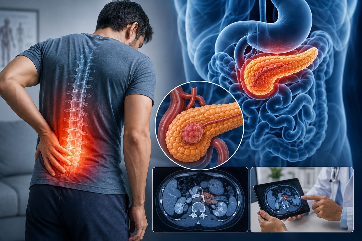
A steady ache rather than a sharp one, often overlooked at first.

Feeling full well before the plate is finished.

Vomiting that follows a heavier, oilier meal in particular.

Dropping weight with nothing in your eating pattern to explain it.
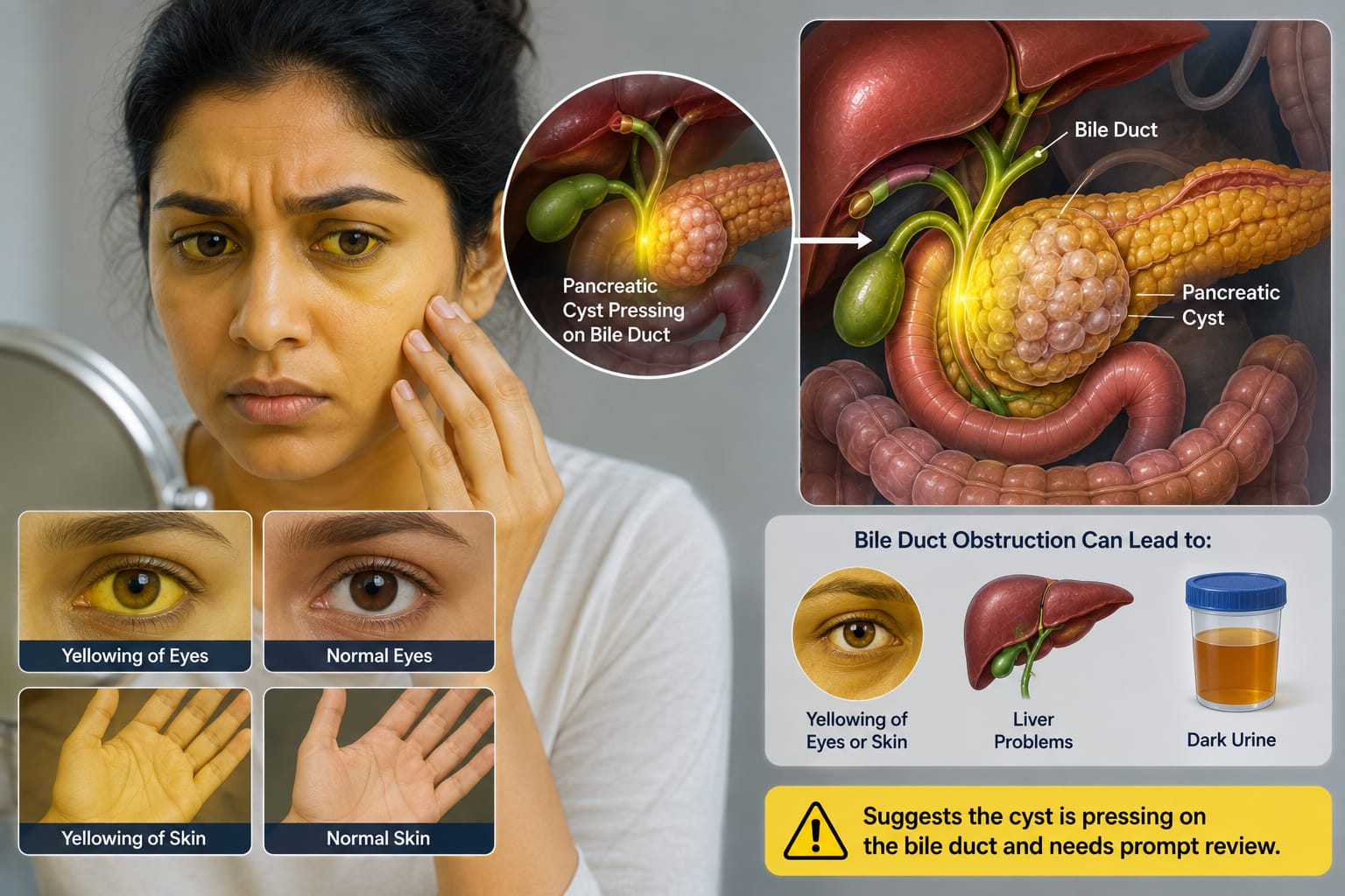
Suggests the cyst is pressing on the bile duct and needs prompt review.

Diabetes without family history, or attacks that keep recurring, both deserve a closer look.
Silence does not mean safety. Certain mucinous cysts enlarge quietly across several years, so one scan alone rarely settles the matter — planned follow-up tracks change over time and answers the question properly.
Causes broadly split into two groups, and knowing the group helps predict behaviour.
A severe acute attack or repeated chronic flares are the most common route to a pseudocyst, alongside blunt injury to the upper abdomen.

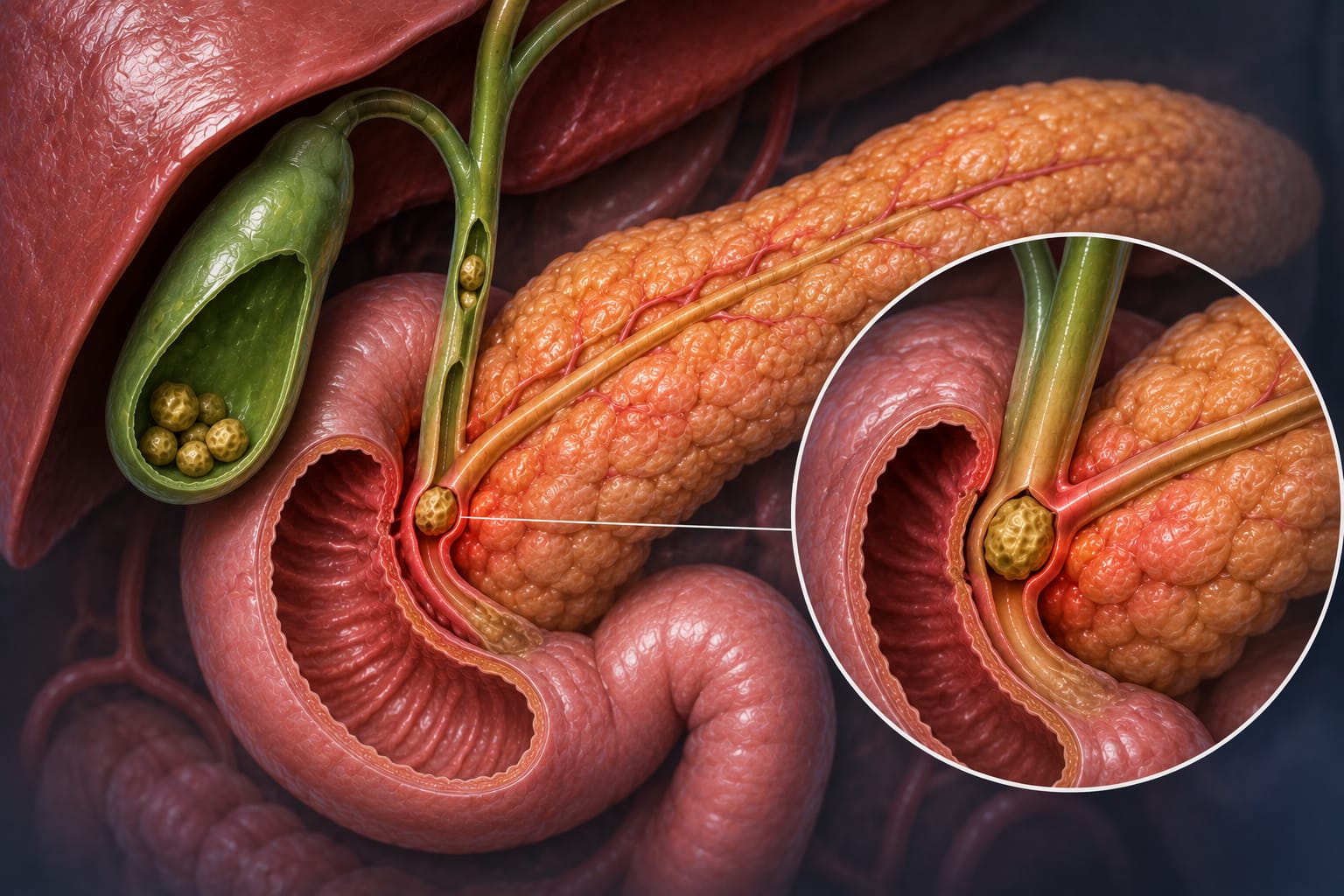
A stone lodged where the pancreatic duct empties can trigger the inflammation that starts a cyst.
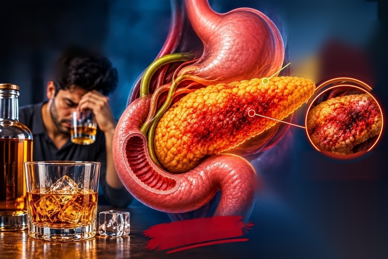
Damages pancreatic tissue directly, and remains one of the leading routes to chronic pancreatitis and, from there, a cyst.

A family history of pancreatic cancer, Von Hippel-Lindau syndrome, or Peutz-Jeghers syndrome all raise the background risk worth mentioning at your first visit.
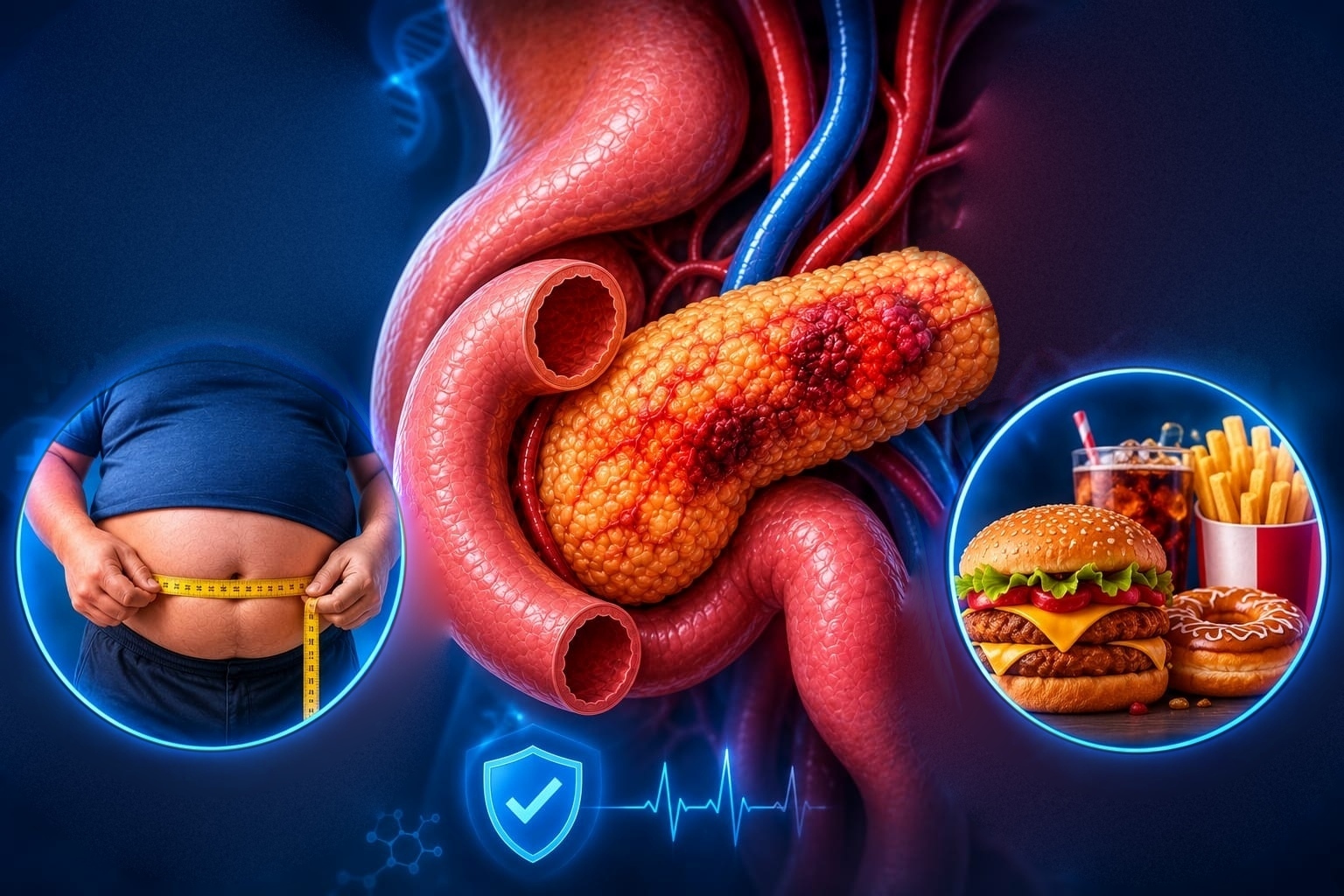
Long-term smoking, along with obesity and poorly controlled diabetes, both raise the odds of a tumour-related cyst developing.
Accurate typing drives every later decision, so we sequence tests deliberately instead of ordering everything at once.
Often finds the cyst first, though it cannot characterise the lesion reliably on its own.
Maps the duct system beautifully without any radiation, and shows whether the cyst connects to the main duct — the single detail that separates IPMN from other types.
A contrast CT scan measures size, detects calcification, and assesses surrounding vessels before any surgical discussion.
Places the probe beside the pancreas through the stomach, dramatically improving resolution, and allows sampling in the same sitting when needed.
CEA, amylase, glucose, cytology and molecular markers on the fluid, alongside HbA1c, LFT and CA 19-9 where relevant — ordered only when the result will actually change the plan.
Treatment follows type, size and symptoms together. Rather than one fixed route, we select from five clear options.
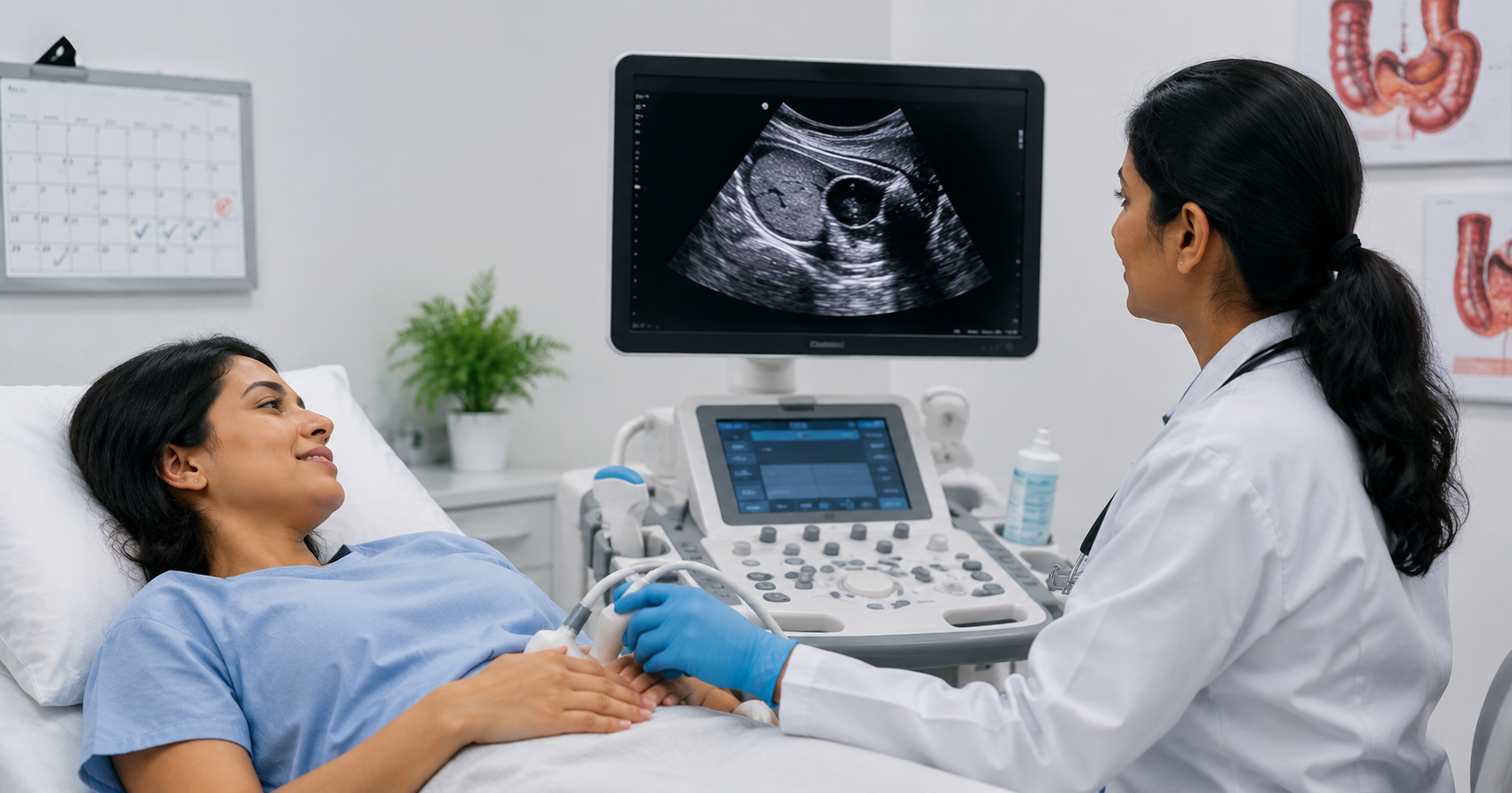
Chosen when the cyst is small, benign-looking and causing no symptoms. Imaging repeats at fixed intervals while you continue normal life without restriction — most patients in our clinic sit in this group.

Chosen when imaging cannot separate a mucinous cyst from a serous one. Under sedation, a fine needle draws fluid while ultrasound guides it, and laboratory analysis settles the type.

Chosen when a mature pseudocyst causes pain, vomiting or infection. The cyst drains into the stomach through a stent instead of open surgery — recovery is quick, with no external cut required.

Chosen when the pancreatic duct or bile duct is blocked. A stent restores flow and relieves jaundice, and settles pain caused by back-pressure at the same time.

Chosen when worrisome or high-risk features appear on scans — main-duct IPMN, mural nodules and rapid growth all qualify. Surgery happens through a hepatobiliary surgical team, and we continue your follow-up afterwards.
Every review checks the same things: change in maximum cyst diameter, main pancreatic duct diameter, any new mural nodule or thickened wall, fresh symptoms such as pain or jaundice, HbA1c, and body weight trend. Five stable years, a small size and a benign type together allow longer gaps between scans — some patients above seventy-five stop imaging altogether after discussion.

Consultant Gastroenterologist & Hepatologist — Gokul Gastro & Liver Care, KPHB · Medicover Hospitals, Financial District.
Dr. Viswanath Kamisetty practises as a pancreas specialist in KPHB and leads Gokul Gastro & Liver Care, holding MBBS, MD and DM in Medical Gastroenterology. Because a single specialist handles your case throughout, nothing gets lost between visits — he personally reviews every scan and every report. Alongside pancreatic cysts, his practice covers acute and chronic pancreatitis, gallstones and bile duct stones, liver disease including fatty liver and cirrhosis, jaundice under evaluation, and gastrointestinal cancers requiring staging.
Pancreatic cystic lesions deserve accurate typing, not anxiety. Most turn out benign and simply need scheduled imaging, while the few high-risk cysts respond well when identified early. Call directly for the earliest slot, or book online, and carry your old reports along.